
Fig. 3.
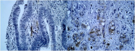
LC3 immunohistochemistry. On the left side, there is a cancer without any dot-like LC3 immunostaining. Notice the positively stained nerve, which can be considered as a positive internal control, since nerve fibers are constantly LC3 positive. On the right side, there is a CRC with a strong and characteristic dot-like LC3 immunostaining. A diffuse cytoplasmic staining was not noticed (×400 magnification)