
Fig. 4.
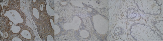
ULK1 immunhistochemistry. On the left, there is a picture of a cancer with strong cytoplasmic immunostaining. In the middle, there is a cancer with barely seen faint ULK1 staining, classified as negative. On the right side, missing staining in normal mucosa is demonstrated. Note the positivity in small blood vessels as an internal control (×400 magnification)