
Fig. 5.
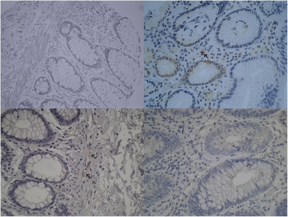
Autophagy-related proteins in normal colonic epithelial tissue. In the upper left, the picture shows normal crypts lacking Beclin-1 immunoexpression. In the upper right, negative p62 staining can be seen in normal colonic crypts. Notice the positive staining in macrophages. The lower left picture demonstrates lack of LC3B expression in crypts. Notice the moderate LC3B positivity in nerve cells. The lower right light micrograph shows a lack of ULK1 staining in normal crypts (×400 magnification)