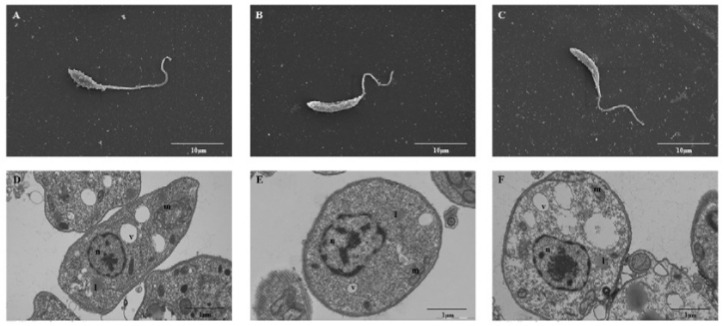
Fig. 2. : ultrastructural analysis of the wild type (WT) and pTEX- or pTEX-heat shock proteins (HSP)70-transfected Leishmania amazonensis promastigotes. Electron microscopy scanning of the A: WT; B: pTEX; and C: pTEX-HSP70 parasites. Ultrathin sections of the D: WT; E: pTEX; and F: pTEX-HSP70 parasites. Abbreviations: l - lipid bodies; m - mitochondria; n - nucleus; v - vacuole. Scale bar 10 µM (A, B and C). Scale bar 1 µM (D, E and F).

