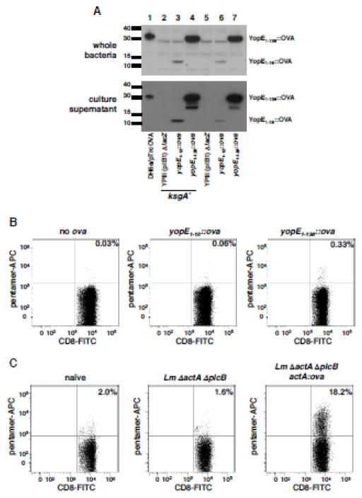
Figure 1. YopE-OVA fusion protein is secreted by Yersinia, and is processed and presented in vivo to stimulate OVA-specific CD8+ T cell responses.
(A) The indicated Y. pseudotuberculosis strains were grown in Yop secretion conditions (Materials and Methods) and processed for western blotting of whole cell lysates and supernatant proteins, with anti-ovalbumin polyclonal antisera. (B) ksgA− strains expressing YopE18::OVA or YopE138::OVA were delivered to animals via intravenous inoculation and 10 days later, spleens removed, single cell suspensions generated and cells incubated with MHC Class I H2-Kb pentamers specific to OVA residues 257–264 and antibodies against CD8 and CD19. Shown are representative histograms showing pentamer and CD8 labeling of CD8+ CD19− cells. (C) Immunization with L. monocytogeneses ΔactA ΔplcB ActA100::OVA generates OVA-specific CD8+ T cells. 3 × 10⁷ CFU of noted L. monocytogeneses strains were delivered to animals via intravenous inoculation. 7 days later spleens were removed, single cell suspensions generated and cells were incubated with H2-Kb OVA257-264 pentamers and antibodies against CD8 and CD19. Shown are representative scatter plots of 2 separated experiments showing pentamer and CD8 labeling of CD8+ CD19− cells (n=3 mice per experiment).

