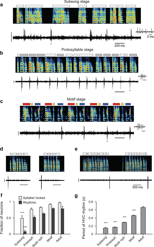
Figure 1. Singing-related firing patterns of HVC projection neurons in juvenile birds.
a, Neuron recorded in the subsong stage, prior to the formation of protosyllables (HVCRA; 51 dph; Bird 7). Top: song spectrogram with syllables indicated above. Bottom: extracellular voltage trace. b, Neuron recorded in the protosyllable stage (HVCRA; 62 dph; Bird 2). Protosyllables indicated (gray bars). c, Neuron recorded after motif formation (HVCRA; 68 dph; Bird 8). d, Neuron bursting exclusively at bout onset (HVCX; 61 dph; Bird 2). e, Neuron bursting exclusively at bout offset (HVCRA; 65 dph; Bird 2). f, Developmental change in the fraction of neurons locked to syllable onsets (gray) and fraction of neurons with rhythmic bursting (black) (mean ± s.e.m; n=39, 135, 566, 378, 32 neurons, respectively). g, Mean period of the HVC rhythmicity as a function of song stage (n=3, 70, 357, 298, 25 neurons, respectively). ***P<0.001, post-hoc comparison with the adult stage. Spectrogram vertical axis 500–8000 Hz. Scale bars: (a–c) 0.5 mV, 200 ms; (d–e) 1 mV, 500 ms. Inset in (a–c) shows zoom of bursts indicated by asterisk; scale bar: 5 ms.

