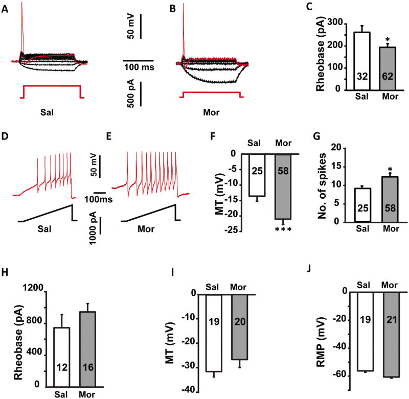
Fig. 1.
Sustained morphine administration for 1 week induced the hyper-excitability of the small diameter neurons (≤ 30 μm) in DRGs, but had no significant effects on larger neurons (> 30 μm). (A, B) Representative recording traces used to measure the rheobase of small neurons from saline and morphine treated rats. (C) Histogram comparing the rheobase in saline (Sal) and morphine (Mor) treated rats. Statistical analysis showed that the rheobase was significant lower in morphine compared to saline treated rats. (D, E) Representative traces used to measure the membrane threshold (MT) in small neurons from saline and morphine treated rats. Membrane threshold (F) was significant lower and the number of spikes (G) was greater for small neurons in morphine compared to saline treated rats. There was no significant difference in rheobase (H), membrane threshold (I) and resting membrane potential (J) in large neurons between saline and morphine treated rats. MT, membrane threshold; RMP, resting membrane potential. * p ≤ 0.05, ***, p ≤ 0.001. Integers in each column represent sample numbers.

