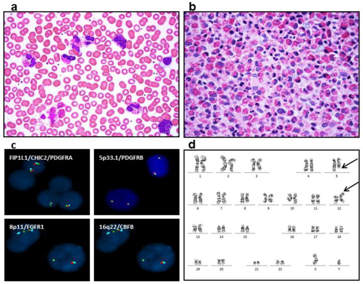
Figure 1.

Marked eosinophilia is detected in patient’s peripheral blood smear (a) and bone marrow core biopsy (b). Fluorescence in situ hybridization analysis (FISH) of rearrangements of FIP1L1/CHIC2/PDGFRA, 5p33.1/PDGFRB, 8p11/FGFR1 and 16q22/CBFB are negative (c). Representative G-banded karyotyping reveals translocation of chromosomes 5 and 12 from peripheral blood cells at metaphases. The breakpoints are identified as 5q31 and 12p13 (arrows) (d).
