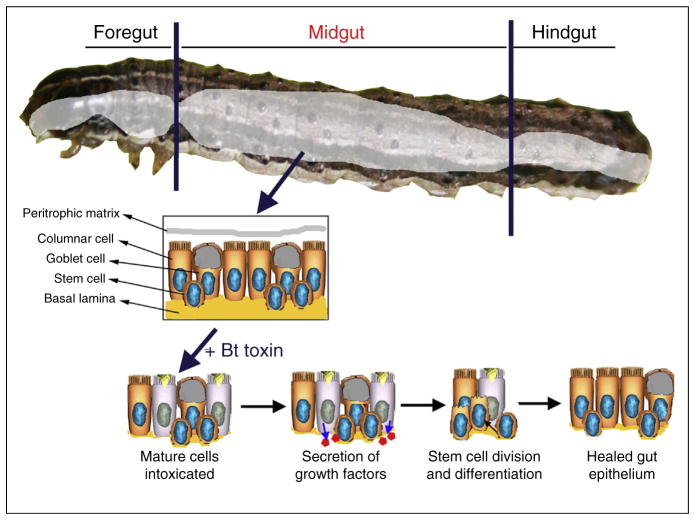
Figure 1.
Diagram of the main cell types in the midgut of lepidopteran larvae and the steps in the process of epithelial healing in response to intoxication with toxins from Bacillus thuringiensis (Bt). Less abundant enteroendocrine cells are also present in the midgut are not represented in the figure.

