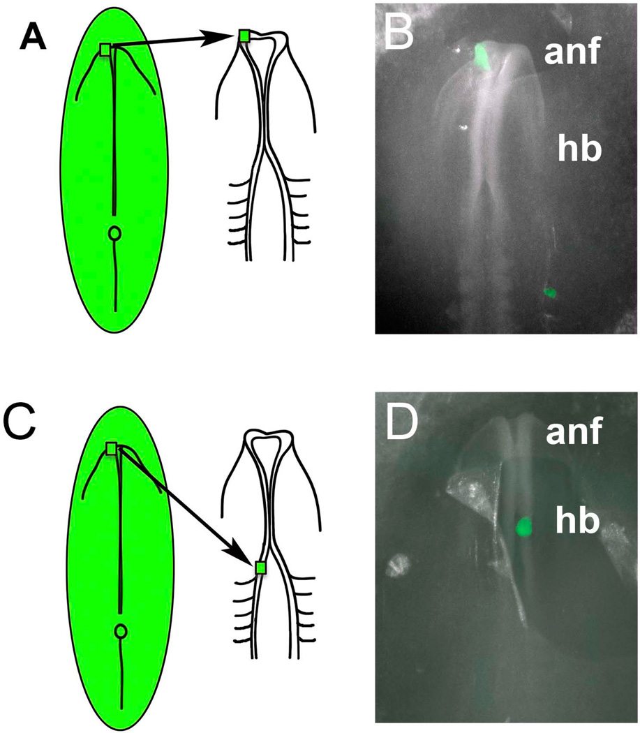
Figure 1.
Schematic diagram illustrating the transplantation paradigm. A) Donor tissue from transgenic GFP chick, transgenic RFP quail or electroporated RFP chick embryos was transplanted from the anterior neural fold (ANF) of the donor embryo into the ANF region of an HH8 host embryo. B) Whole mount image of an embryo after a GFP-transgenic chick graft (green) was transplanted isotopically into the left side of an unlabelled host embryo. C) Donor tissue from transgenic GFP chick, transgenic RFP quail or electroporated RFP chick embryos was transplanted from the anterior neural fold (ANF) of the donor embryo into the rostral hindbrain (HB) region of an HH8 host embryo. D) Whole mount image of an embryo after a GFP-transgenic chick graft (green) was transplanted heterotopically into the right side of an unlabelled host embryo.

