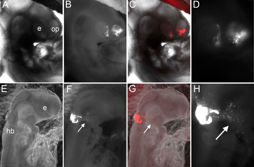
Figure 2.
Results of anterior neural fold (ANF) to ANF transplants (A–D) and ANF to rostral hindbrain (rHB) transplants (E–H) after two days of incubations. A) Bright field, (B, C) Bright field plus fluorescence, and Fluorescence channel alone at higher magnifcation (D) of an embryo two days after grafting an RFP-electroporated ANF in place of the ANF of unlabeled host. Labeled cells populate the olfactory placode epithelium (OP) as well as the eye (E). E) Bright field, (F,G) Bright field plus fluorescence, and Fluorescence channel alone at higher magnification (H) of an embryo two days after grafting an RFP-electroporated ANF into the rostral hindbrain (HB) of an unlabeled host. Labeled cells populate contribute to the hindbrain and also migrate away, as expected for neural crest cells.

