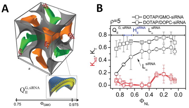
Fig. 3.
Double gyroid cubic phase for the delivery of siRNA (A) The unit cell of the cubic phase contains a negative Gaussian surface of lipids (grey) separating 2 water channels that contain siRNA (orange and green). (B) Both lamellar complexes (LαsiRNA, circles) and cubic complexes (QIIG, siRNA, squares) show low non-specific silencing (red curves) at low membrane charge density but the cubic phase significantly out performs the lamellar phase at total gene knockdown (black curves). Reprinted with permission from (35). Copyright 2010 American Chemical Society.

