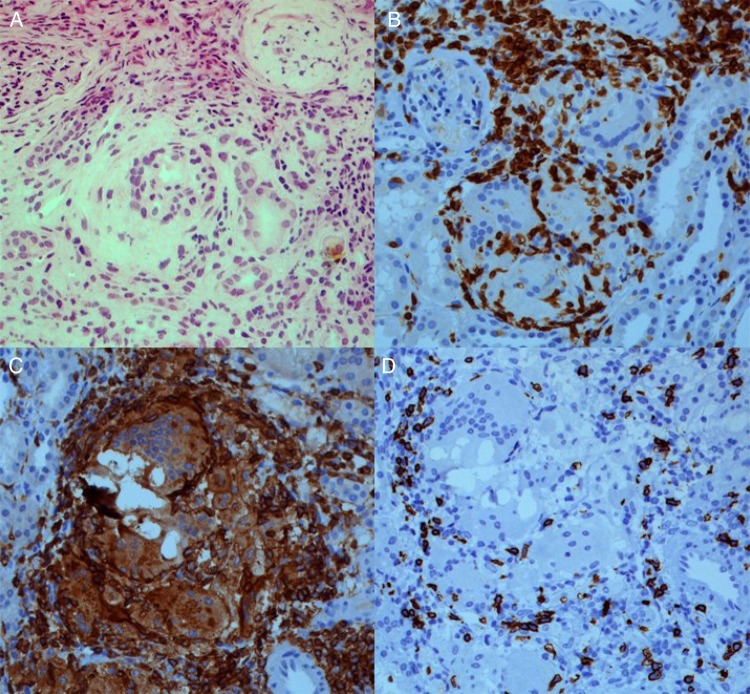
Fig. 1.
Renal biopsy revealing granulomatous interstitial nephritis (A; haematoxylin and eosin, 200×). Immunohistochemistry staining revealing a predominant T cell infiltrate (B; CD3, 200×), consisting of both T-helper cells (C; CD4, 200×) and cytotoxic T cells (D; CD8, 200×). Of note, neither crystals nor polarized material was observed.

