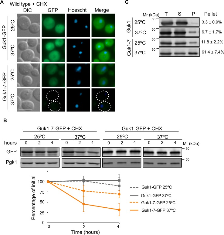
Fig 2. Misfolded Guk1-7 is degraded at the non-permissive temperature.
(A) Wild type cells expressing ectopic Guk1-GFP or Guk1-7-GFP were grown at 25°C and then incubated in the presence of the translation inhibitor cycloheximide (CHX) at 25°C or 37°C for 2 hours prior to fixation and imaging. Scale bar represents 5μm. (B) Cycloheximide chase assay. Wild type cells expressing ectopic Guk1-GFP or Guk1-7-GFP were incubated with CHX for 4 hours at 25°C or 37°C and samples were collected at the indicated time points. Guk1-GFP and Guk1-7-GFP was immunoblotted with anti-GFP antibodies and a representative blot is shown. GFP levels were normalized to Pgk1 levels and shown in the graph below with results representing the means and standard deviations of three independent experiments. (C) Guk1-GFP and Guk1-7-GFP were ectopically expressed in wild type cells grown at 25°C or shifted to 37°C for 20 min. Total cell lysate (T), soluble (S), and pellet fractions (P) were immunoblotted with anti-GFP antibodies. The ratio of the pellet fraction to total cell lysate is noted and represents the mean and standard deviation of three independent experiments.

