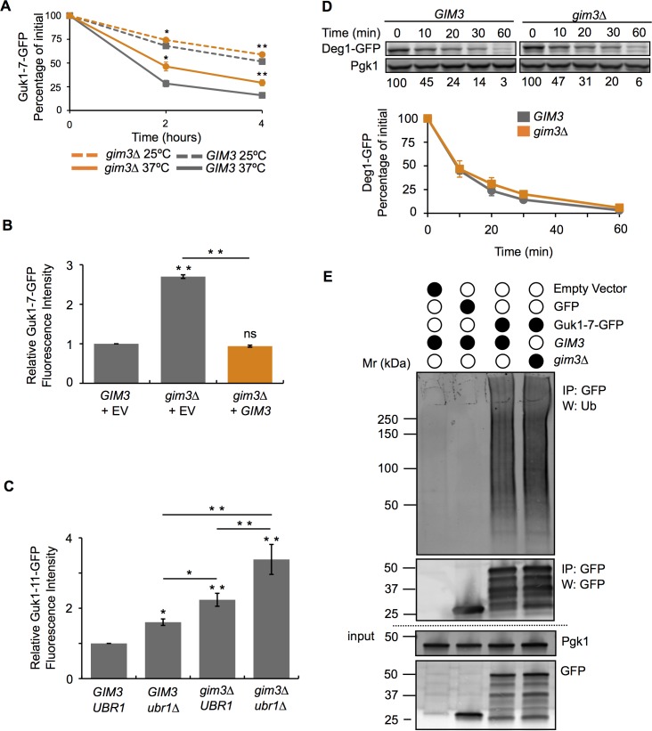
Fig 6. Absence of Gim3 reduces Guk1-7 turnover.
(A) Wild type and gim3∆ cells expressing Guk1-7-GFP were incubated with CHX at 25°C or 37°C and samples were analysed by flow cytometry at the indicated time points. The results represent the means and standard deviations of three independent experiments and the asterisk denotes significance of P < 0.05. (B) Gim3 addback experiment. GIM3 or gim3∆ cells expressing Guk1-7-GFP and either an empty vector (EV) control or GIM3. The results represent the means and standard deviations of three independent experiments of the relative fluorescence intensity after a two hour CHX incubation at 37°C. P values were calculated with a one-way ANOVA and post-hoc Tukey HSD to assess significance, ** denotes P < 0.005. (C) Wild type, ubr1∆, gim3∆, and ubr1∆gim3∆ cells expressing Guk1 (T290G) fused to GFP were incubated at 37°C with CHX for two hours and then analysed by flow cytometry. P values were calculated with a one-way ANOVA and Holm multiple comparison to assess significance, * and ** denote P < 0.05 and 0.01, respectively. (D) Proteasome activity assay. Gim3 or gim3∆ cells expressing Deg1-GFP under the Cup1 promoter were incubated at 30°C in the presence of CHX and samples were collected at the indicated time points. Deg1-GFP was immunoblotted with an anti-GFP antibody. The results represent the mean and standard deviation of three independent experiments. (E) GIM3 or gim3∆ cells expressing Guk1-7-GFP, GFP alone, or a control empty vector were grown at 25°C. Guk1-7-GFP was immunoprecipitated with GFP-Trap beads and eluted samples were immunoblotted with anti-ubiquitin and anti-GFP antibodies.

