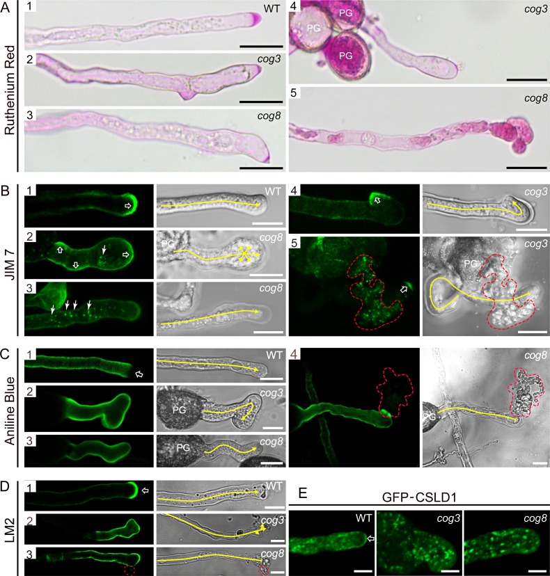
Fig 8. Altered distribution of cell wall materials and proteins in cog3 and cog8 pollen tubes.
(A) Ruthenium red staining of pollen tubes. The crescent region in wild-type pollen tubes was stained heavily by ruthenium red (A1) and the deformed tips of cog3 and cog8 pollen tubes showed less staining (A2, A3, and A4). A bursting pollen tube with strong ruthenium red staining was trapped inside the cytosol (A5). (B) Confocal and corresponding DIC images of wild-type (B1), cog3 (B4, B5), and cog8 (B2, B3) pollen tubes labeled with JIM7 monoclonal antibody. Open arrow in (B1) indicates the crescent region of a wild-type pollen tube, while open arrows in (B2), (B4), and (B5) indicate the bulging tips of a curved pollen tube. Arrows in (B2) and (B3) indicate JIM7-positive structures inside cog3 and cog8 pollen tubes. (C) Confocal and corresponding DIC images of wild-type (C1), cog3 (C2), or cog8 (C3, C4) pollen tubes stained with aniline blue. Open arrow in (C1) indicates the tip region of a wild-type pollen tube. (D) Confocal and corresponding DIC images of wild-type (D1), cog3 (D2), and cog8 (D3) pollen tubes labeled with LM2 antibody. LM2 stained the very tip region (indicated by an open arrow) of wild-type pollen tubes. The tip-localization pattern of the LM2 signal was absent in cog3 and cog8 pollen tubes. (E) Confocal images of wild-type (E1), cog3 (E2), and cog8 (E3) pollen tubes expressing GFP-CSLD1. Red dashed lines in (B5), (C4), and (D3) highlight the cytoplasmic outflow of ruptured cog3 and cog8 pollen tubes. Yellow lines in (B), (C), and (D) indicate the pollen tube growth direction. Bars = 20 μm in (A); 10 μm in (B), (C), and (D); 5 μm in (E).

