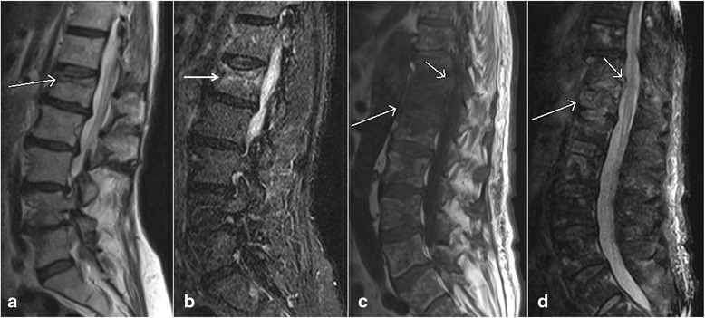
Fig. 12.

Sagittal T1 weighted image (a) and sagittal T2 weighted image (b) show a compression fracture of L1 vertebral body with a horizontal band of bone marrow edema in the superior half of vertebra without associated soft tissue mass, suggesting a benign compression fracture. Sagittal T1 weighted image (c) and sagittal T2 weighted image (d) of a different patient with multiple metastases show compression fractures with diffuse involvement of vertebral body and associated epidural soft tissue mass. Note abnormal bone marrow signal intensities also seen in other vertebral bodies, reflecting diffuse metastatic disease
