Fig. 4.

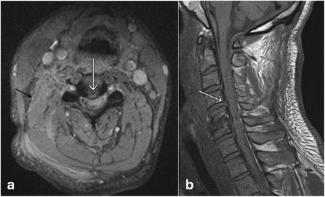
Axial gradient recalled echo (GRE) image (a) and sagittal T1 weighted image (b) show the presence of a small central disc herniation (white arrows). Also note the presence of paraspinal muscle edema (black arrow, a)

Axial gradient recalled echo (GRE) image (a) and sagittal T1 weighted image (b) show the presence of a small central disc herniation (white arrows). Also note the presence of paraspinal muscle edema (black arrow, a)