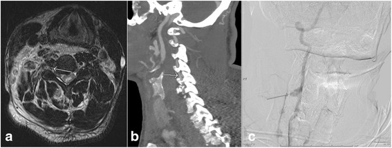
Fig. 6.

Axial T2 weighted image (a) shows the presence of post traumatic vertebral artery dissection with double lumen (arrow). Subsequent CT angiogram of the neck (b) confirms of the finding of vertebral artery injury (arrow). Follow-up angiography of the neck performed on the next day (c) shows the presence of pseudoaneurysm (arrow)
