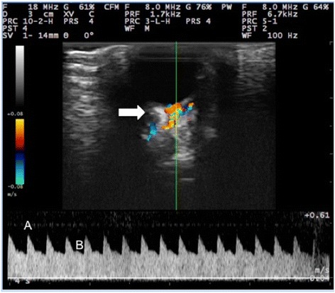
Fig. 1.

Color Doppler image and pulsed wave flow of the external ophthalmic artery of an adult male New Zealand white rabbit treated with 10 mg of sildenafil citrate (day 15). Blood flow in the external ophthalmic artery towards the transducer shown in red and flow in the opposite direction in blue. The external ophthalmic artery exhibited a laminar flow pattern with intermediate resistivity, dichroism, and two peak systolic velocities (a and b). Optic nerve (white arrow)
