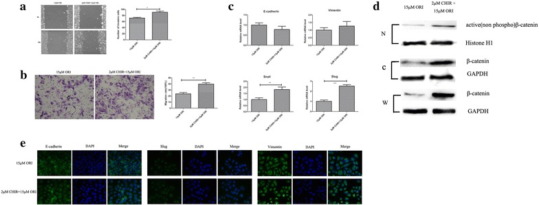
Fig. 4.

CHIR attenuated the inhibitory function of ORI. a and b Cell migration was determined by using wound healing and transwell assay. The migrative ability of CHIR group (2 μM CHIR + 15 μM ORI) was compared with that seen in the ORI group (15 μM). c Real-time PCR analysis of EMT markers were compared between CHIR group(2 μM CHIR + 15 μM ORI) and ORI group (15 μM). d Representative western blots for active β-catenin and β-catenin(total) levels in ORI or CHIR group. Histone H1 and GAPDH were used as internal control. (N, nucleus; C, cytoplasm; W, whole cell). e Immunofluorescence of EMT markers were compared between CHIR group (2 μM CHIR + 15 μM ORI) and ORI group (15 μM). Data are presented as the mean ± SD, n = 3. *P < 0.05, **P < 0.01, ***P < 0.001 vs. ORI group (15 μM)
