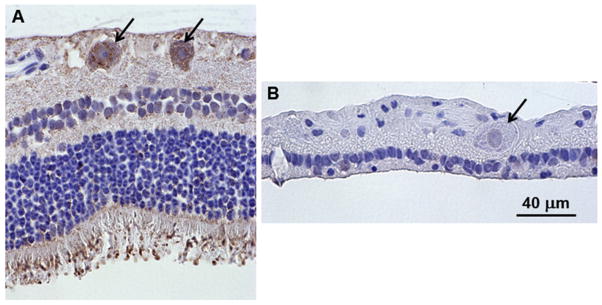
Fig. 6.
Light micrographs of retinal cross-sections immunostained with an anti-TPP1 antibody. The brown color indicates TPP1 antibody binding. (A) The retina from a normal Dachshund shows punctate immunostaining in the ganglion cells (arrows in A). (B) Ganglion cells from Dog B did not show any TPP1 immunolabeling (arrow in B). Intense TPP1 immunostaining was also present around the photoreceptor outer segments in the normal dog and diffuse immunostaining was present throughout most of the rest of the retina. Both retinas were artifactually detached from the retinal pigment epithelium during processing for paraffin embedding. Bar in (B) indicates the magnification of both micrographs.

