
Fig. 3. Unbiased epidermal fate tracking by single-cell photolabeling.
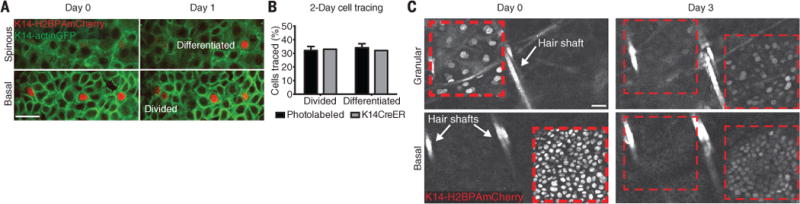
(A) Representative examples of cell division and differentiation fates. (B) Quantification of cell division and differentiation events. (C) Representative time sequence of a region labeled with a photoactivatable reporter. At day 0, two adjacent square areas were scanned to activate the H2BPAmCherry reporter in the granular and basal layer, respectively. Scale bars, 25 μm.