FIG 2.
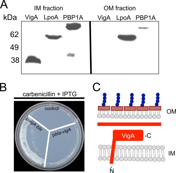
VigA is an inner membrane protein. (A) Cells carrying either ectopically expressed, His-tagged VigA or chromosomally encoded, His-tagged PBP1A (control protein for inner membrane localization) or LpoA (control protein for outer membrane localization) were lysed and separated into fractions via differential centrifugation. Proteins were visualized by immunoblotting with an antibody recognizing the His epitope. OM, outer membrane fractions; IM, inner membrane fractions. (B) Ectopically expressed N- or C-terminal fusions of VigA with beta-lactamase were plated onto agar containing an inducer (IPTG, 100 μM) and carbenicillin. The control is an empty plasmid. bla, beta-lactamase. (C) Model of VigA topology based on the experiments described above. C, C terminus.

