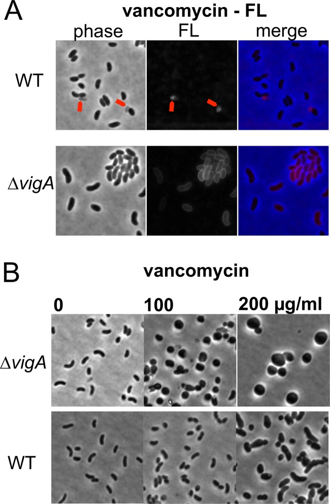
FIG 3.
VigA mutant cells appear more permeable to vancomycin than do WT cells. (A) Cells were grown in LB medium, washed once in PBS, stained for 10 min with fluorescent vancomycin (vancomycin FL), washed twice with PBS, and then imaged. The red arrowheads point to lysed WT cells. (B) Prior to imaging, cells were grown to exponential phase (optical density at 600 nm of 0.5) in LB medium and then exposed to the indicated concentrations of vancomycin for 3 h.

