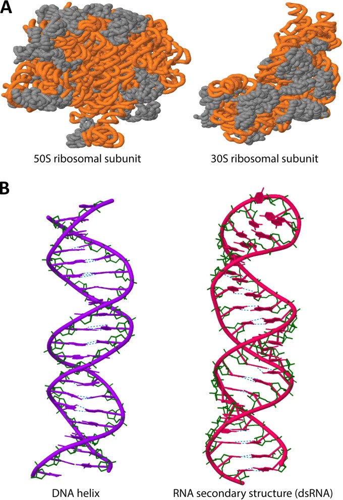
FIG 1.
(A) Structures of the 50S and 30S bacterial ribosomal subunits, showing the secondary structures of rRNA (orange) in association with ribosomal proteins (gray). (B) Structures of dsDNA and dsRNA, showing hydrogen bonds between bases (blue) and other intramolecular bonds (green). Structure templates were obtained from the Protein Data Bank (PDB) and drawn using Jmol (an open-source Java viewer for chemical structures in three dimensions).

