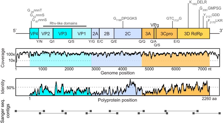
FIG 2 .
Poecivirus genome organization. (Top) Predicted genome organization. P1 (blue) represents viral structural proteins, and P2 (violet) and P3 (orange) represent nonstructural proteins. Predicted N-terminal cleavage sites are shown below the bar, and conserved picornaviral amino acid motifs are shown above it. (Middle) Number of reads from the metagenomic sequencing data set that support each base. (Bottom) Polyprotein homology between poecivirus and its closest relative, duck megrivirus, measured as the pairwise identity of a moving 15-amino-acid window.

