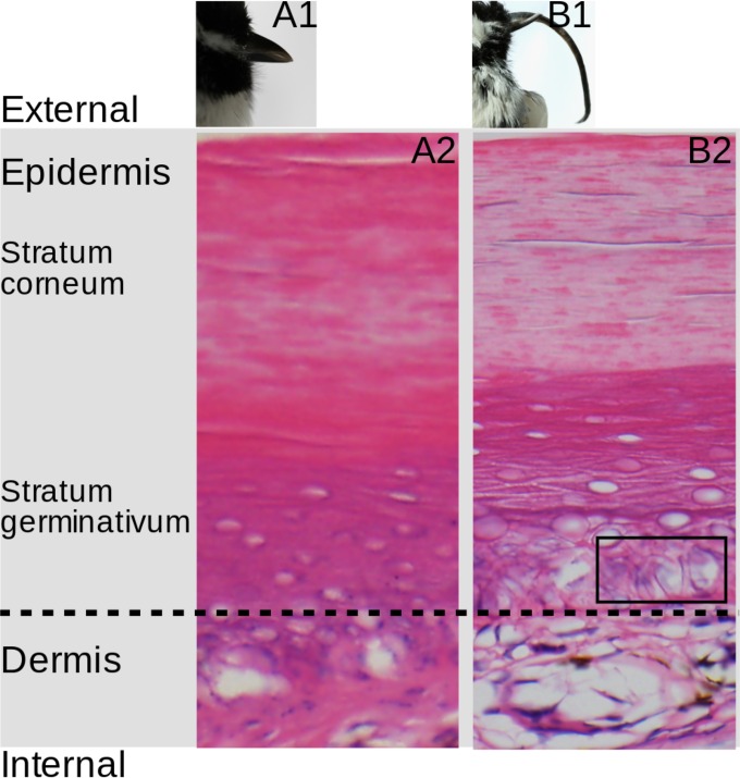
FIG 5 .
Histopathology of AKD. (A1 and B1) Gross beak morphology of BCCH 601 (unaffected by AKD) and 498 (AKD affected; beak length is 40.3 mm), respectively. (A2 and B2) Histopathology of the beaks of BCCH 601 (A2) and 498 (B2). The black box (B2) indicates an area of cytoplasmic vacuolization of cells; the nuclei of these cells show contour irregularities and areas surrounded by a rim of clear cytoplasm, creating an “owl eye” appearance.

