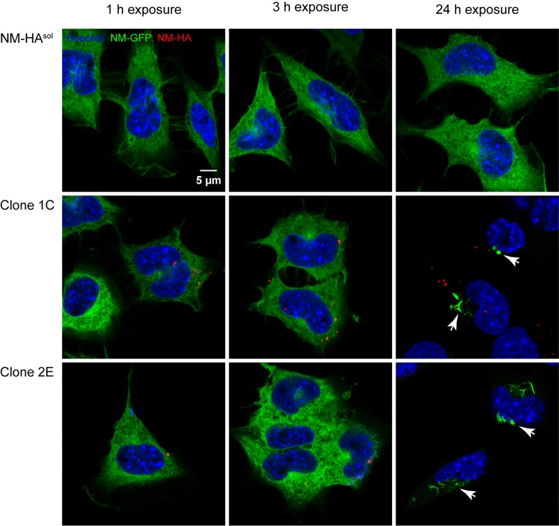
FIG 1 .
Murine N2a cells harboring cytosolic Sup35 NM-HA aggregates secrete infectious NM prions. Shown are confocal images of recipient N2a NM-GFP cells (green) exposed to the 100,000 × g pelleted fractions derived from conditioned medium of NM-HA aggregate-producing cell clones 2E and 1C or control donor cells expressing soluble NM-HA. Recipient cells were fixed 1, 3, and 24 h posttreatment with pellet fractions. NM-GFP aggregate induction was observed after 24 h with pellet fractions derived from media of both clones but not from donor cells expressing NM-HAsol. NM-HA was stained with anti-HA antibody (red), and nuclei were counterstained with Hoechst (blue). NM-GFP aggregates are indicated by arrows. For images showing cells with NM-GFP aggregates, the microscopy setting had to be adjusted to prevent the overexposure of highly fluorescent aggregates. Note that no colocalization of internalized NM-HA seeds and NM-GFP aggregates was observed. Scale bar, 5 µm.

