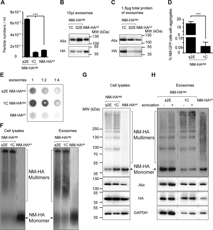
FIG 5 .
Aggregation state of NM-HA associated with exosomes. (A) Vesicle concentrations in the P4 fractions derived from media of NM-HAagg clones s2E and 1C and NM-HAsol cells measured by ZetaView nanoparticle tracking analysis. The results shown are means ± SD (n = 3; ***, P < 0.001; one-way ANOVA). (B, C) Western blot analyses of P4 fractions loaded at comparable volumes of isolated exosomes or adjusted to comparable total protein levels. The positions of the lanes were switched for presentation purposes (dashed lines). (D) Cell-based aggregate induction assay. Percentages of recipient cells with NM-GFPagg induced by P4 exosomal fractions of donor clones 1C and s2E are shown. The results shown are means ± SD (n = 6; ***, P < 0.0001; unpaired Student t test). (E) Filter trap assay using P4 fractions isolated from clones s2E and 1C and NM-HAsol control cells. Shown at the top are the dilutions used. (F) SDD-AGE analysis of cell lysates and exosome fractions derived from NM-HAsol cells and NM-HAagg clones s2E and 1C. (G, H) Glutaraldehyde cross-linking with cell lysates and exosomes from NM-HAsol cells and clones s2E and 1C (top). The same amount of sample without cross-linking was also loaded as a control and analyzed for Alix, HA, and GAPDH protein levels (bottom). Extra marker lanes were removed for presentation (dashed lines).

