FIG 1 .
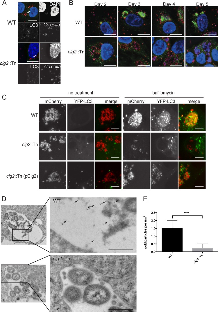
LC3 is abundant in the CCV lumen. (A) Fluorescence micrographs of HeLa cells 24 h after infection with either the parental strain of C. burnetii (WT) or the isogenic cig2::Tn mutant. Merged panels show staining for LC3 (green), Coxiella (red), and DAPI (blue). Bars, 10 µm. (B) Fluorescence micrographs of HeLa cells infected with either the parental strain of C. burnetii (WT) or the cig2::Tn mutant at the indicated times after infection. Panels show staining for LC3 (green), Coxiella (red), and DAPI (blue). Bars, 10 µm. (C) HeLa cells producing YFP-LC3 were infected for 5 days with the indicated strains of C. burnetii producing mCherry. Live-cell imaging was used to visualize mCherry (red) and YFP-LC3 (green) in cells that were either left untreated (no treatment) or treated with bafilomycin for 1 h prior to imaging. Bars, 5 µm. (D) Images obtained by cryo-immunoelectron microscopy of HeLa cells producing GFP-RFP-LC3 infected with the indicated C. burnetii strains for 5 days. Arrows highlight the locations of anti-GFP-labeled gold particles that colocalized with GFP-RFP-LC3 associated with autophagic bodies in the lumen of vacuoles formed by C. burnetii (WT). Bars, 500 nm. (E) The density of gold particles in the lumen of the CCV was determined for 10 different vacuoles and plotted as the average number of gold particles per square micrometer. ****, P < 0.0001.

