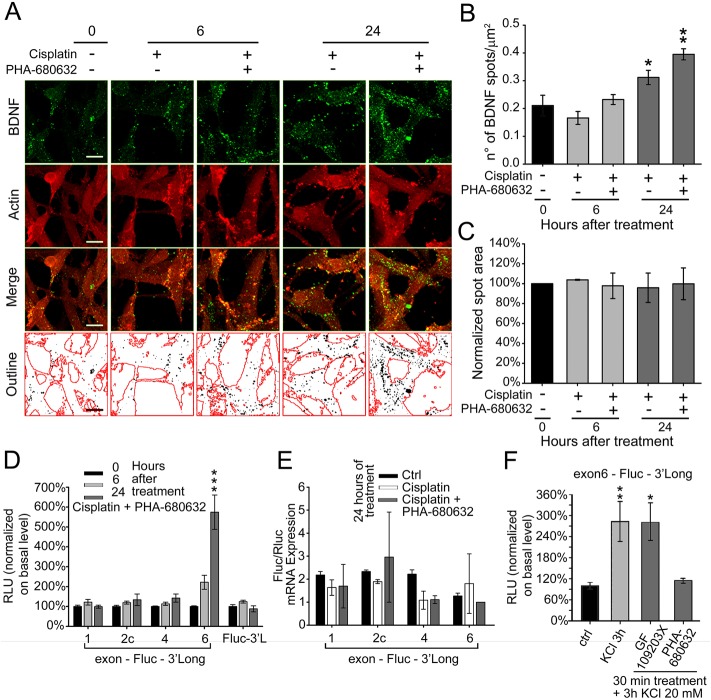
Fig. 4.
Increment in BDNF protein production and exon-6 translation upon combined treatment of cisplatin and PHA-680632. (A) Immunofluorescence on SK-N-BE cells treated with cisplatin±PHA-680632 (aurora inhibitor) at different time points: untreated control (0), 6 and 24 h. BDNF spots are detected using a mouse mAb and actin is marked using a rabbit pAb to emphasize cell bodies; the merge of the two channels is shown as well as its outline, where black dots represent the BDNF spots and red lines the edges of the cell bodies (N=3; scale bar: 10 µm). (B) BDNF spot number analysis from the immunofluorescence described in panel C. Data are given as number of spots per µm2. N=3; *P=0.039, **P=0.004; one-way ANOVA versus untreated) (C) BDNF spot area analysis, normalized to untreated control. (D) Translational capacity of different BDNF isoforms in differentiated SK-N-BE treated as described in panel A. The isoforms tested and the normalization procedures are the same as described in Fig. 2, panel C. RLU, relative luciferase unit. N=3, each in duplicate. ***P<0.001; one-way ANOVA untreated control). (E) Quantitative real-time PCR to evaluate the effects of treatment, for 24 h with cisplatin±PHA-680632, on the expression of firefly luciferase (Fluc) mRNA, normalized to the expression of the Renilla luciferase used as transfection control (N=2). (F) Effect on exon 6-Fluc-3′Long efficiency of translation after 20 mM KCl treatment for 3 h, alone or in combination with GF 109203X (PKC inhibitor) or PHA-680632 (aurora inhibitor) for 30 min of treatment. N≥3; *P=0.03, **P=0.006; one-way ANOVA versus control). For all panels, data are given as mean±s.e.m.

