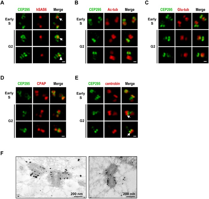
Fig. 2.
Super-resolution (3D-SIM) analysis of the subcellular localizations of CEP295 and other centriolar proteins during centriole biogenesis. U2OS cells were synchronized by aphidicolin for 24 h (to arrest cells at early S phase) and the synchronized cells were released in fresh medium for another 8 h (to allow progression to G2 phase). The cells were then fixed and dual stained with antibodies against CEP295 (A–E) and hSAS6 (A), Ac-tub (B), glutamylated tubulin (Glu-tub) (C), CPAP (D) or centrobin (E). Super-resolution images were acquired using a Zeiss ELYRA system. (A) Arrows indicate that CEP295 embraces hSAS6 at the proximal end of the procentriole, arrowhead indicates two-dot shape with hSAS6 in the center. (E) Arrows indicate localization of centrobin distal to CEP295 on growing centrioles. Scale bars: 0.2 μm. (F) Immunogold electron microscopy analysis of CEP295 localization.

