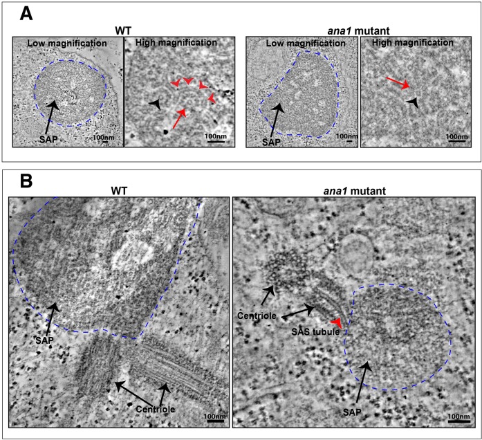
Fig. 3.
Ana1 is not essential for SAP assembly. (A) Micrographs show low- and high-magnification views from electron microscopy tomographs of a SAP particle (dotted blue line) in a WT (left panels) or ana1 mutant (right panels) spermatocyte. The SAP in the WT cell is highly structured and consists of well-organised ‘SAStubules’ that resemble the central cartwheel of the centriole (Stevens et al., 2010b). An end-on view of a SAStubule is highlighted by the black arrowhead, and a transverse view is highlighted with red arrowheads. The SAStubules are linked by spokes to an electron-dense outer ring (red arrow). In ana1 mutants, the SAP structures are less well-defined, but the central cartwheel-like SAStubules are still easily discerned (red arrow), especially in an end-on view (black arrowhead). (B) The SAPs in WT spermatocytes are often directly connected to the centrioles, and we could detect small centriole-like structures associated with the SAPs in ana1 mutants in seven out of 11 of the SAPs we examined: in this example the central cartwheel of one of the centriole-like structures appears to be directly connected to a SAStubule that extends into the SAP (red arrowhead).

