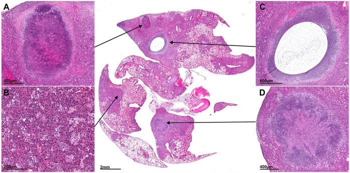
Fig. 3.
Lesion heterogeneity. Histopathological section from the lung of a representative M. tuberculosis-infected (H37Rv, 14 wpi; without TB treatment) mouse is shown. Higher-power views (insets) demonstrate multiple pathologies – granulomas with central caseation (A,D), pneumonia (B) and cavitation (C) – in different areas of the same lung tissue simultaneously.

