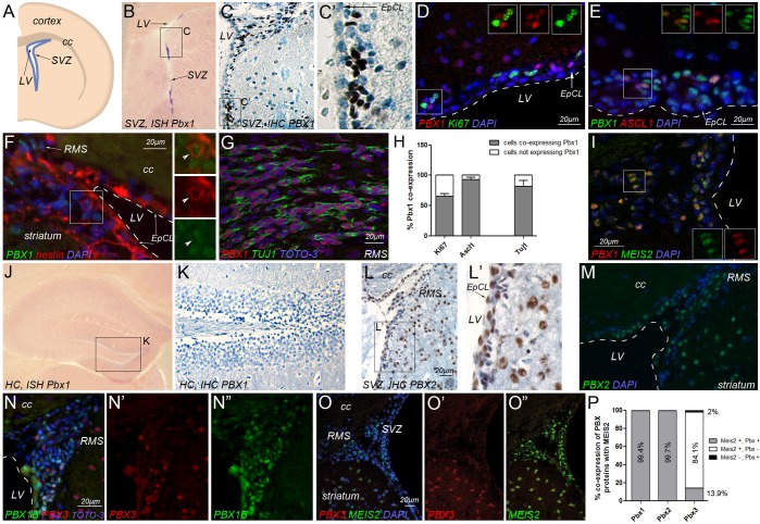
Fig. 1.
PBX expression in the SVZ. (A) Schematic representation of the adult mouse SVZ. (B) In situ hybridization for Pbx1 transcripts (blue) in the SVZ. (C) PBX1 protein (brown) in the SVZ; cell nuclei are counterstained with Hematoxylin (blue). The boxed area is shown at higher magnification in C′. (D-F) Immunofluorescence double labeling for (D) PBX1 (red) and Ki67 (green), (E) PBX1 (green) and ASCL1 (red) or (F) PBX1 (green) and nestin (red). Boxed areas are shown as single channels. Arrow (D) marks the EpCL; arrowhead (F) marks a representative nestin+/PBX1+ cell located adjacent to the EpCL. (G) PBX1 (red) protein in migrating TUJ1+ neuroblasts (green) in the RMS. (H) Quantification of colocalization of PBX1 with Ki67, ASCL1 or TUJ1. Error bars indicate s.e.m. (I) PBX1 (red) and MEIS2 (green) in neuroblasts at the beginning of the RMS. (J,K) Lack of Pbx1 transcript (J) and protein (K) in the hippocampus. (L-M) PBX2 protein in the SVZ and RMS. Ependymal and striatal cells are strongly immunoreactive for PBX2, whereas cells in the subependyma are weakly immunoreactive for PBX2. The boxed region is magnified in L′. (N-O″) Double-stainings reveal limited overlap between PBX3 (red) and (N-N″) PBX1B (green) or (O-O″) MEIS2 (green) in the SVZ. The PBX1B antibody was used for double immunofluorescence labeling with PBX3 or MEIS2 (both rabbit polyclonal antibodies). PBX1B is the prominent PBX1 isoform expressed in SVZ-derived stem/progenitor cells (Fig. S6). (P) Quantification of colocalization of MEIS2 with different PBX family proteins in the SVZ and emerging RMS. n=2 (E,F,N) or n=4 (D,I,O), hemispheres. cc, corpus callosum; EpCL, ependymal cell layer; HC, hippocampus; IHC, immunohistochemistry; ISH, in situ hybridization; LV, lateral ventricle; RMS, rostral migratory stream; SVZ, subventricular zone.

