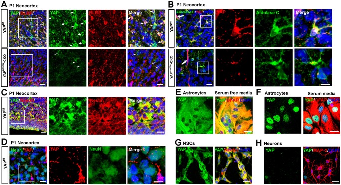
Fig. 1.
Selective expression of YAP in neocortical NSCs and astrocytes. (A) Double immunostaining of YAP (green) and BLBP (red) in the layer III-V neocortex of P1 Yapf/f and Yapnestin-CKO mice. Arrows and arrowheads indicate typical YAP+/BLBP+ and YAP+/BLBP− cells, respectively. The boxed region is magnified to the right as single-channel and merged images. (B) Double immunostaining of YAP (red) and aldolase C (green) in the neocortex of P1 Yapf/f and Yapnestin-CKO mice. Arrows indicate the blood vessels. (C,D) Double immunostaining of (C) YAP (green) and nestin (red) and (D) YAP (red) and NeuN (green) in the neocortex of P1 Yapf/f mice. (E,F) Double immunostaining of YAP (green) and GFAP (red) in primary cultured astrocytes with (E) serum-free medium or (F) 10% FBS+DMEM. (G,H) Double immunostaining of (G) YAP (green) and nestin (red) in primary cultured NSCs or (H) YAP (green) and MAP2 (red) in primary cultured neocortical neurons (4 days in vitro) from WT neocortex. DAPI (blue) was used to stain nuclei. The YAP antibody used in B and D was from CST (#8418/D24E4), otherwise from Sigma (WH0010413M1). Scale bars: 20 μm.

