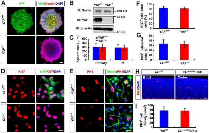
Fig. 2.
Normal proliferation and self-renewal of NSCs in Yapnestin-CKO mice. (A) Double immunostaining of YAP (green) and nestin (red) in cultured neurospheres from E14.5 Yapf/f and Yapnestin-CKO mice. (B) Western blot analysis of YAP expression in WT and YAP-deficient neurospheres. IB, immunoblot antibody. (C) Quantification of WT and YAP-deficient neurosphere size in primary cultures or the fifth passage of the primary cultures (n=100 per group). (D,E) Double immunostaining analysis of (D) Ki67 (red) and YAP (green) or (E) PH3 (red) and nestin (green) in NSCs from WT and YAP-deficient neurospheres. (F,G) Quantitative analysis of the percentage of (F) Ki67+ cells (n=45 fields per group) and (G) PH3+ cells (n=30 fields per group) among total WT or YAP-deficient NSCs. (H) Immunostaining analysis of PH3 in the neocortex of E14.5 Yapf/f and Yapnestin-CKO mice. (I) Quantitative analysis of PH3+ cells in Yapf/f and Yapnestin-CKO mice (n=8 sections in WT group, n=6 sections in mutant group). DAPI (blue) was used to stain nuclei. Data are mean±s.d. Scale bars: 20 μm.

