
Fig. 2.
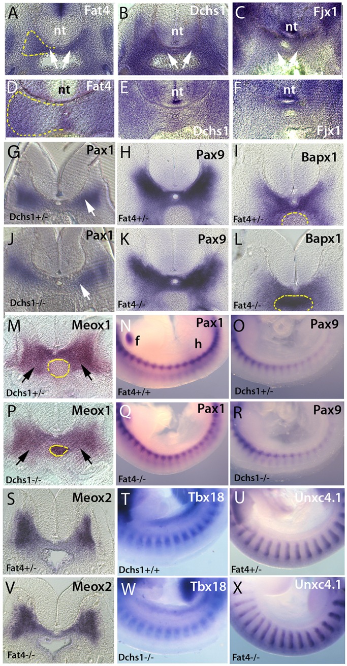
Sclerotome specification is unaffected in Fat4 and Dchs1 mutants. In situ hybridisation showing expression of Fat4 (A,D), Dchs1 (B,E) and Fjx1 (C,F) in the E10.5 (A-C) and E11.5 (D-F) wild-type sclerotome (arrows, A-C). (G-X) Transverse sections (G-L,M,P,S,V) and lateral views (N,O,Q,R,T,U,W,X) of in situ hybridisations of E10.5 wild-type/heterozygous (G-I,M-O,S-U), Dchs1−/− (J,P,R,W) and Fat4−/− (K,L,Q,V,X) embryos showing expression of the indicated genes. Arrows (G,J,M,P) indicate the developing sclerotome. Dashed yellow lines outline one half of the developing vertebral body to show the region of the sclerotome analysed for cell proliferation and apoptosis (A,D) or the aorta (I,L,M,P) to show the sclerotomal expression more clearly. In N, the forelimb (f) and hindlimb (h) have been labelled to indicate the orientation of the embryos in lateral views. nt, neural tube.