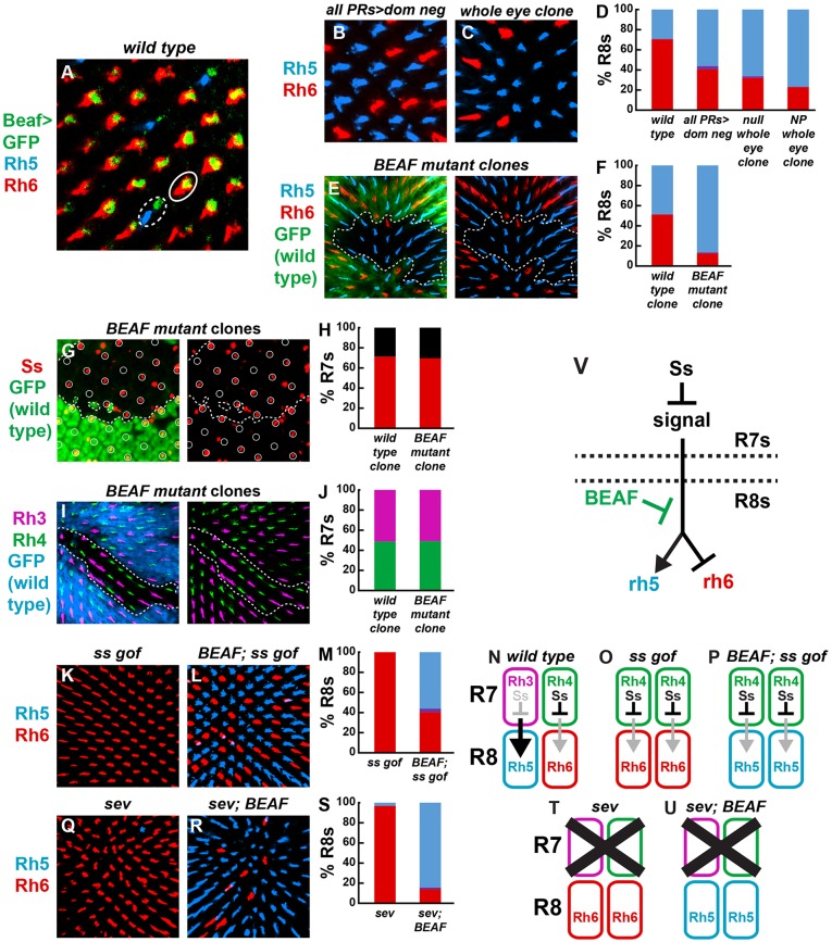
Fig. 2.
BEAF acts in R8s downstream of R7 signaling to control Rhodopsins. (A) BEAF-GFP under control of the BEAF promoter was expressed in both R8 subtypes. Rh5-expressing pR8 (dashed oval); Rh6-expressing yR8 (solid oval). (B) Rh5-expressing R8s increased and Rh6-expressing R8s decreased when a BEAF dominant-negative construct was expressed specifically in photoreceptors. (C) A similar phenotype was observed in whole eye BEAF null mutant clones. (D) Quantification of the data shown in B,C. (E,F) BEAF null mutant clones (GFP−) contained more R8s expressing Rh5 compared with wild-type or heterozygous tissue (GFP+). Dashed lines represent clone boundary in all panels unless otherwise noted. (G,H) Ss was expressed stochastically with similar frequency in BEAF null (GFP−) and control (GFP+) tissue in pupal retinas. R7 cells are circled. Red indicates percentage of R7s expressing Ss; black indicates percentage of R7s lacking Ss. (I,J) The Rh3 and Rh4 expression ratio was normal in BEAF null mutant clones (GFP−). (K) Ectopic Ss expression in all R7s induced Rh6 and inhibited Rh5 expression in nearly all R8s. (L) Ectopic Ss expression in the absence of BEAF resulted in increased Rh5- and decreased Rh6-expressing R8s. (M) Quantification of the data shown in K,L. (N-P) Schematics depicting wild type, K and L. (Q) Genetic ablation of R7s (and hence the signal to R8) in sev mutants caused expression of Rh6 and loss of Rh5 in nearly all R8s. (R) sev; BEAF null double mutants displayed upregulation of Rh5 and downregulation of Rh6. (S) Quantification of the data shown in Q,R. (T,U) Schematics describing the observations shown in Q,R. (V) Model for how BEAF acts in R8s, downstream of R7 signaling to control Rhodopsins.

