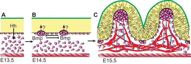
Fig. 3.
Villus formation and patterning in the mouse. (A) The intestinal epithelium (yellow) is pseudostratified (not shown); both apical (green) and basal (black) epithelial surfaces are flat at E13.5. The epithelium secretes Hh ligands that signal in a paracrine manner to underlying mesenchymal cells (pink). The intestine is encircled by the inner circular muscle (red hatching). (B) At E14.5, Hh signals trigger mesenchymal cells to cluster, beginning in the anterior small intestine. A self-organizing Turing-type field drives cluster patterning via mesenchymal Bmp signaling. Clusters send unknown signals (indicated by ‘?’) back to overlying epithelial cells, causing them to shorten. (C) Progressive shape changes in epithelial cells cause villus emergence at each cluster by E15.5. New clusters form beneath the still-proliferative intervillus epithelium, directing another round of villus emergence. Vasculature (red) and ectodermally derived nerves (blue) connect to the clusters. The inner circular muscle (red hatching) may confine the epithelium, promoting rapid villus emergence. Throughout this time, Pdgfa is expressed by the epithelium and Pdgfra is expressed by mesenchymal clusters. Disruption of this signaling pathway reduces subsequent rounds of villus formation, but does not perturb emergence of the first villi.

