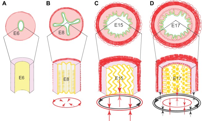
Fig. 4.
Villus formation and patterning in the chick. (A-D) Schematic representations of intestinal cross-sections (top) and partial longitudinal views (bottom) illustrate sequential development of patterned epithelial folds that precede villus patterning. (A) At E6, prior to visceral muscle development, a loose mesenchyme (pink) surrounds the epithelium (yellow); the epithelial apical surface (green) is flat. (B) By E8, the outer circular muscle forms, confining the proliferative epithelium and leading to development of longitudinal ridges. (C) Formation of the outer longitudinal muscle at E15 bends the ridges into zigzags. (D) Formation of the inner longitudinal muscle is concomitant with villus emergence. Red arrows indicate the direction of compression conveyed by the addition of each muscle layer; black arrows show forces imposed by previously formed muscles. The progressive muscle-induced epithelial bending creates localized pockets of concentrated Hh signals that drives patterned mesenchymal cluster formation and villus emergence.

