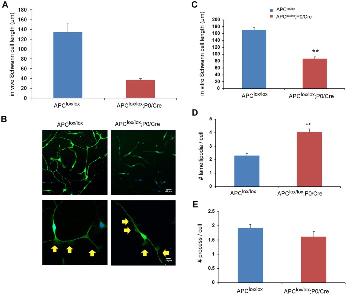
Fig. 6.
Loss of APC results in perturbed Schwann cell process extension and lamellipodia formation. 3DEM images of serial sections were acquired from sciatic nerves of Apclox/lox;P0-Cre and Apclox/lox (control) mice at P7 and individual Schwann cells traced and analyzed in both genotypes. (A) Detailed analysis of a single Schwann cell in the 3DEM images of sciatic nerves revealed the presence of shorter cell processes in Apclox/lox;P0-Cre mice. (B) Primary Schwann cells were cultured and stained for S100, a Schwann cell marker. The Schwann cells derived from the Apclox/lox;P0-Cre sciatic nerves were much shorter (quantified results in C). Increased numbers of lamellipodia per cell were detected in the Schwann cells derived from the Apclox/lox;P0-Cre sciatic nerves (quantified results in D). Yellow arrows indicate lamellipodia. (E) No significant changes in the number of cell processes were found in the mutant cells compared with controls. Number of Schwann cells traced and analyzed in vivo in the sciatic nerves: Apclox/lox, n=5; Apclox/lox;P0-Cre, n=27; **P<0.001. Number of cells analyzed in vitro (primary culture): Apclox/lox, n=414; Apclox/lox;P0-Cre, n=469; **P<0.001.

