Fig. 3.
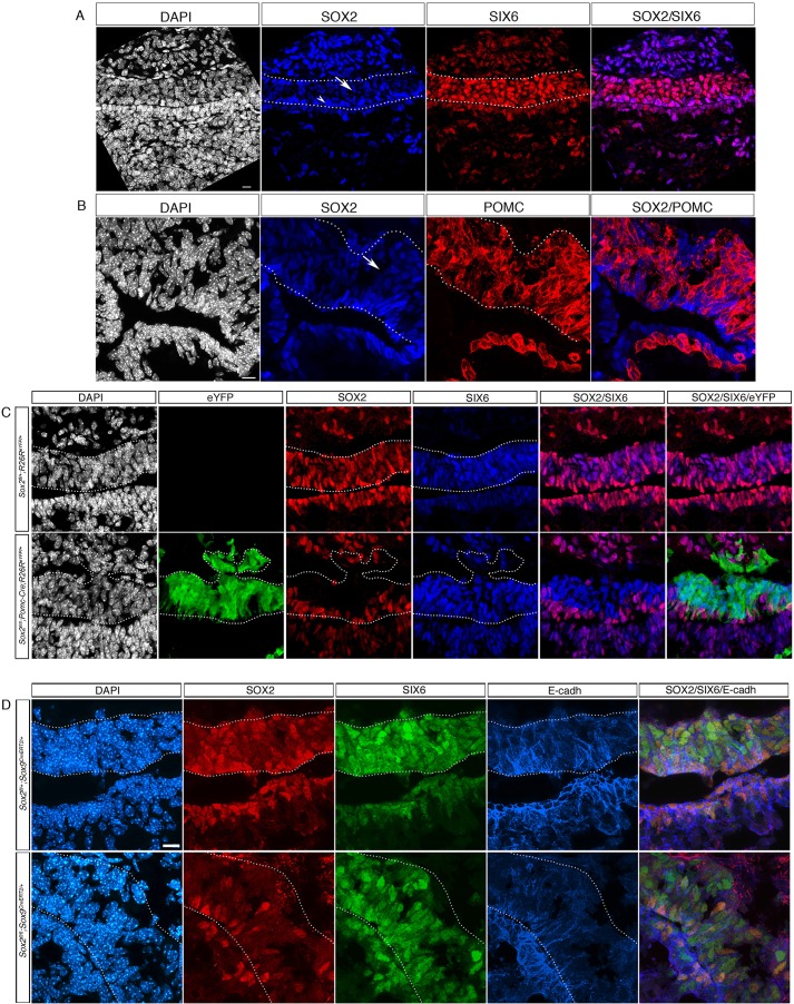
SOX2 regulates SIX6 expression exclusively in the SOX2Hi progenitor population. (A) Immunofluorescence for SOX2 and SIX6 at 18.5 dpc in a control embryo. SOX2 is highly expressed in cells lining the pituitary lumen (SOX2Hi; arrowhead), and at lower levels in cells within IL (SOX2Low; arrow). Both SOX2-positive cell populations uniformly express SIX6. (B) Immunofluorescence for SOX2 and POMC at 18.5 dpc in a control embryo. SOX2Low cells are POMC-positive melanotrophs (arrow). (C) Immunofluorescence for SOX2, SIX6 and eYFP in a Pomc-Cre;Sox2fl/fl;R26ReYFP/+ embryo at 18.5 dpc. SOX2Low expression is specifically lost in eYFP-positive melanotrophs, whereas eYFP-negative, SOX2Hi cells are maintained around the cleft. SIX6 expression in IL is unaffected by the loss of SOX2. (D) Immunofluorescence for SOX2, SIX6 and E-cadherin in Sox2fl/+;Sox9CreErt2/+ and Sox2fl/fl;Sox9CreErt2/+ embryos at 18.5 dpc, induced by tamoxifen at 13.5 dpc. SOX2 expression is lost in some E-cadherin-positive cells in the epithelial cell layer lining the cleft. Deletion of SOX2 does not result in downregulation of SIX6. Scale bar: 10 μm in A,B,D; 5 μm in C. IL is outlined.

