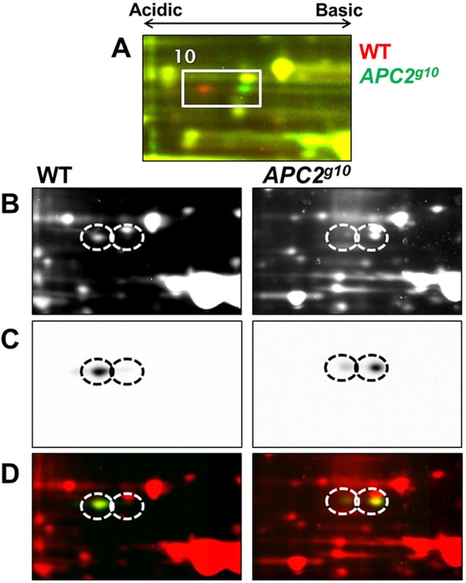
Fig. 4.

Immunoblot confirmation that CaBP1 is an APC2-dependent difference-protein. (A) 2D-DIGE image of the region containing CaBP1 isoforms. (B) Fluorescent images of wild-type and Apc2g10 embryo lysates that were separately labeled with Cy3 DIGE dye, resolved on different 2DE gels and transferred to nitrocellulose. (C) Immunoblot images of proteins stained with anti-CaBP1 antibody. (D) Superimposed images of total Cy3-labeled protein (red) and the CaBP1 2D immunoblot (green) confirm protein identification by LC-MS/MS. The predominant isoform found in wild-type lysate is the left isoform, whereas the right isoform is predominant in Apc2 null embryos.
