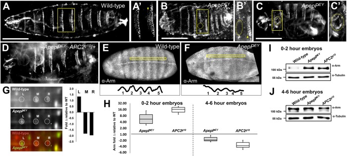
Fig. 7.
ApepPEY embryos display Wnt activation phenotypes similar to Apc2 mutant embryos. (A-F) Anterior is to the left and posterior is to the right. (A-C′) Wild-type (A) and ApepPEY mutant (B,C) cuticles. ApepPEY cuticles exhibit head defects, fusion of the first and second denticle rows (compare A′ with B′, arrowhead) and denticle loss (B′, circled). ApepPEY cuticles also have body holes (C,C′, circled). (D) Dose reduction of Apc2 in ApepPEY embryos modifies the ApepPEY phenotype. This example has head defects and denticle loss. (E,F) Wild-type (E) Arm protein localization and accumulation reflect patterned Wnt pathway activation, whereas ApepPEY mutant embryos (F) exhibit a more uniform accumulation of Arm protein, as exemplified in the traces beneath. (G) 2D-DIGE comparison of wild-type and ApepPEY embryos showing that total ApepP protein is decreased in mutant embryos and that there is a shift in isoform distribution toward the left isoform in ApepPEY embryos. (H-J) Anti-Arm immunoblots of embryo lysates at 0-2 h (I) and 4-6 h (J) AEL. At 0-2 h AEL Arm significantly accumulates in both mutants, consistent with reduced Arm degradation, whereas at 4-6 h AEL Arm levels are decreased in both mutants (H; n=3 replicates).

