Fig. 2.
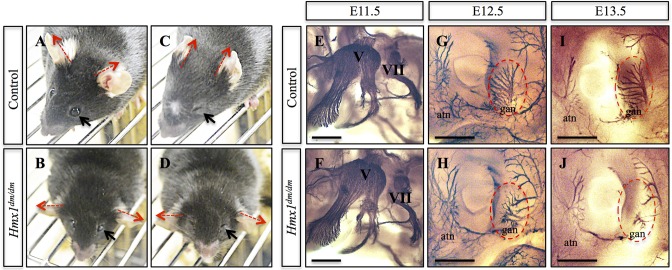
Hmx1dm/dm animals show abnormal response to an aversive facial stimulus and defects in sensory nerve branches. (A-D) Representative images of control (A,C) and Hmx1dm/dm (B,D) mice before (A,B) and during the air puff test (C,D) demonstrate that Hmx1dm/dm mice, in contrast to controls, do not move their ears in response to having air blown on their face (compare red dashed arrows in A with C, and B with D; control n=8, Hmx1dm/dm n=7), although Hmx1dm/dm mice can effectively close their eyes (compare black arrow in A with C, and B with D). (E,F) Neurofilament staining of E11.5 embryos using a 2H3 antibody demonstrate that the trigeminal (V) ganglion is intact in Hmx1dm/dm embryos (control n=6, Hmx1dm/dm n=5). (G-J) Noticeable defects can be seen by 2H3 staining in the great auricular nerve (gan) and the auriculotemporal nerve (atn) that surround and innervate the pinna of Hmx1dm/dm embryos at both E12.5 (compare G with H, red dashed oval; control n=6, Hmx1dm/dm n=4) and E13.5 (compare I with J, red dashed oval; control n=8, Hmx1dm/dm n=2). Scale bars: 500 µm (E-J).

