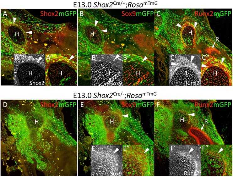
Fig. 2.
Deletion of Shox2 results in specific loss of Shox2+/Runx2+ perichondrial cells. (A-F), Co-immunofluorescence of Shox2, mGFP, Sox9 and Runx2 in the forelimb of Shox2Cre/−;RosamTmG embryos at E13.0 (D-F) compared with littermate Shox2Cre/+;RosamTmG mice (A-C) shows loss of distinct stylopodial perichondrial structure (arrowheads) in the absence of Shox2 compared with that of zeugopod (arrow in F). Humerus regions are magnified in inserts A′,A″,B′,B″,C′,C″,E′,E″,F′,F″. H, humerus; R, radius.

