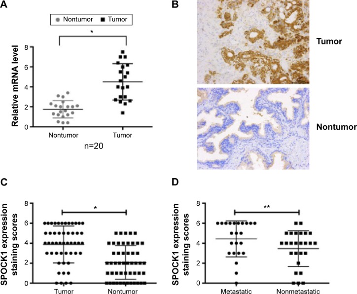
Figure 1.
SPOCK1 is overexpressed in prostate cancer tissues.
Notes: (A) qRT-PCR analysis of SPOCK1 in 20 cases of prostate cancer. Levels of SPOCK1 in tumor and the adjacent nontumor tissues were detected and compared. (B) IHC analysis of the protein expression of SPOCK1 in 50 cases of prostate cancer. Representative images showing the high staining of SPOCK1 in tumor tissues were shown. (C) After the scoring of IHC staining, all the 50 tumor cases and 50 nontumor cases were classified into each score. Staining scores of SPOCK1 in the tumor tissues were significantly higher than the nontumor tissues. (D) The 50 cases were divided into those with metastasis (n=24) and those without (n=26). It was further shown by IHC that the average staining score of SPOCK1 in metastatic tissues was significantly higher than the nonmetastatic tissues. *P<0.001; **P<0.05, as indicated.
Abbreviations: SPOCK1, SPARC/osteonectin, cwcv, and kazal-like domain proteoglycan 1; IHC, immunohistochemistry analysis; qRT-PCR, quantitative real-time polymerase chain reaction.

