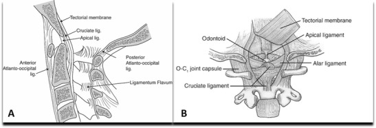
Fig. 1.

Artistic illustration of lateral (a) and posterior (b) views of the craniovertebral junction and the stabilizing ligaments. Note that the cruciate ligament is composed of horizontal fibers (i.e., transverse ligament) and vertical fibers. b The tectorial membrane, the rostral extent of the posterior longitudinal ligament, has been reflected in (b) to allow for visualization of more ventral structures
